
Escucha gratis este podcast en la aplicación: Descarga gratuita en la App Store
radio.net
Sleep timer
Favoritos
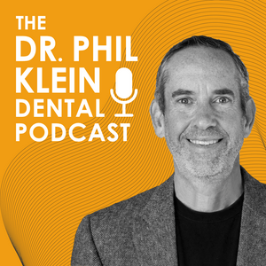
The Dr. Phil Klein Dental Podcast
Dr. Phil Klein | Dentist
Último episodio
809 episodios
Ep. 814 - The Right Perio Mix for the GP
22/10/2026 | 36 minAre you confident that your hygiene department is truly diagnosing and treating periodontal disease at the level your patients deserve — or are you leaving significant clinical and financial opportunity untreated?
Dr. Marc Goldman is a board-certified periodontist with over 40 years of clinical experience, founder of a premier periodontal practice in Millburn, New Jersey, and an educator at a general practice residency program at Beth Israel Hospital in Newark. With decades of mentoring general dentists and residents on the realities of periodontal and implant therapy, Dr. Goldman brings both the clinical depth and the practice management perspective that few specialists can offer.
This episode examines one of the most underappreciated revenue and risk management opportunities in general dentistry: periodontal therapy. Dr. Goldman and Dr. Phil Klein break down which periodontal services GPs should confidently keep in-house, which cases warrant referral, and how the GP-periodontist relationship should function as a collaborative business partnership rather than a competitive one. The conversation covers crown lengthening, implant treatment planning, hygiene department efficiency, and the real-world consequences of underdiagnosing periodontal disease — including malpractice exposure.
Episode Highlights:
Scaling and root planing is consistently underutilized in general practices where hygienists perform prophylaxis on patients who clinically require debridement at a therapeutic level. Using local anesthesia to anesthetize one or two quadrants at a time dramatically improves patient comfort, clinician efficiency, and case throughput, with per-quadrant fees substantially exceeding standard prophylaxis reimbursement.
The full-mouth debridement code is frequently misused as a preliminary step before diagnosis, but this approach wastes a clinical visit, reduces reimbursement, and displaces the opportunity to initiate scaling and root planing immediately. If periodontal disease is present and the patient requires root planing and scaling, initiating treatment by quadrant during that same appointment is both more efficient and more appropriate.
Crown lengthening is a significantly underutilized procedure in general practice, partly due to misconceptions about post-operative healing timelines. In many cases, adequate gingival healing occurs within two to three weeks, allowing the referring dentist to take a final impression or adjust a provisional restoration shortly thereafter — making the workflow disruption far less significant than commonly perceived.
Implant treatment planning requires a systematic, multi-specialty approach before any surgical intervention is initiated. Key pre-implant considerations include periodontal health, attached tissue adequacy, bone volume and proximity to anatomical structures such as the sinus, adjacent tooth vitality, and whether orthodontic tooth movement is indicated to optimize implant site dimensions.
Underdiagnosing or under-presenting periodontal disease carries both malpractice risk and significant consequences for long-term patient retention. Patients who later discover untreated moderate or advanced periodontal disease through a second provider consistently lose trust in their original dentist, resulting in permanent loss of that patient relationship and potential legal exposure.
Perfect for: general dentists at any career stage who want to optimize their periodontal and hygiene department profitability, dental residents building foundational practice management skills, and any clinician looking to develop stronger specialist referral relationships and reduce medico-legal risk.
If you want a clearer picture of where your practice stands on periodontal diagnosis, treatment, and referral strategy, this conversation will give you the framework to act on it.Ep. 813 - The Last Steps in Indirect Restorations Make All the Difference
19/10/2026 | 41 minAre you treating every indirect restoration the same way at cementation — and wondering why some cases still fail or leave patients sensitive? The answer often comes down to a few critical chairside decisions that most dentists make on autopilot.
Dr. Jennifer Derse is a restorative and cosmetic dentist with advanced training in clear aligner therapy and indirect restorations. A graduate of Marquette University School of Dentistry, she has lectured internationally and serves as a mentor at the Kois Center in Seattle, Washington — one of the most respected postgraduate dental education institutions in the world. Dr. Derse also leads clinical mentorship for incoming doctors at Espire Dental and has completed executive education programs at Harvard Business School Online, McKinsey, and the Ritz Carlton Leadership Institute.
In this episode, Dr. Derse walks through her complete cementation protocol for lithium disilicate and zirconia restorations, explaining how material choice should drive every downstream clinical decision — from intaglio surface preparation and bonding agent selection to cement choice and isolation strategy. The conversation is grounded in Kois Center-trained principles and enriched by Dr. Derse's hands-on mentorship experience watching real clinicians make real mistakes. Dr. Phil Klein, an endodontist and DMD, contributes additional clinical depth on dentin tubule anatomy and pulp proximity that every restorative dentist should understand before bonding near the pulp.
Episode Highlights:
Lithium disilicate and zirconia require completely separate preparation protocols for the intaglio surface. Air abrasion is contraindicated on glass-ceramic restorations, while hydrofluoric acid etching is contraindicated on zirconia. Confusing these two tracks is one of the most common cementation errors seen in mentored clinical settings.
For lithium disilicate crowns with an enamel margin, combining air abrasion with selective phosphoric acid etch on enamel produces superior mechanical retention compared to acid alone. The universal bonding agent is then scrubbed into both enamel and dentin — with the self-etching chemistry of the adhesive handling dentin bonding without the risks of total-etch technique.
Silane must be applied as a standalone agent, not as a component embedded within a bonding agent. Because universal bonding agents are acidic — with pH values around 3.2 — they destabilize silane over time. For adhesively retained restorations with limited ferrule, a separate silane application step is essential to preserving the chemical bond at the ceramic-cement interface.
For veneer cementation, light-cure resin cement offers superior control compared to dual-cure formulations because the ultra-thin restoration allows full light penetration. Try-in paste — a non-adhesive, shade-matched glycerin gel — allows the clinician and patient to preview the final color effect before committing to cement shade, which is especially important when masking dark underlying tooth structure or achieving very high-value aesthetics.
Post-cementation sensitivity is often preventable with two key steps: using a dual-cure resin cement that integrates into dentinal tubules as its pH neutralizes during placement, and applying a calcium- and fluoride-containing self-adhesive liner when the preparation approaches the pulp. The anatomy of dentin itself matters — intertubular dentin decreases significantly near the pulp as tubule diameter increases, reducing available bonding surface and making pulp protection even more critical in deep preparations.
Perfect for: General dentists and restorative specialists who place indirect restorations, dental residents building their cementation protocols, and any clinician who wants to stop treating lithium disilicate and zirconia the same way at the cementation appointment.
If you have ever had a patient return with sensitivity, a dislodged veneer, or a shade result that surprised you, this episode gives you the clinical framework to make sure it does not happen again.Ep. 812 - What Burnout Really Looks Like in Dentistry and How to Reset
15/10/2026 | 40 minAre the tough days getting harder to shake, or is something deeper building beneath the surface of your clinical life?
Tiffany Castellanos is a licensed clinical social worker (LCSW) and practicing psychotherapist with a master's degree in social work. With a prosthodontist husband and close ties to the dental community, she brings a uniquely informed perspective to the psychological demands of dental practice. She works with dentists virtually, including in early-morning sessions designed to fit clinical schedules, and is currently collaborating with the University of Florida College of Dentistry to develop a continuing education curriculum on dentist well-being.
This episode examines the psychological undercurrents of dental burnout—what it looks like before it becomes a crisis, and what dentists can realistically do about it during and after the clinical day. Drawing on both psychotherapy research and firsthand exposure to the dental profession, Tiffany offers a framework grounded in nervous system regulation, cognitive behavioral principles, and leadership communication. The conversation is especially timely given that over half of dentists will experience some form of burnout, 40% report considering leaving the profession, and dentists carry a suicide rate two to three times higher than the general population.
Episode Highlights:
Burnout is defined as a state in which prolonged, chronic work-related stress causes job demands to exceed a person's emotional, physical, and mental capacity. Early warning signs include an inability to recharge even after adequate sleep, increased irritability and reduced empathy toward patients, and a shift from professional satisfaction to simply getting through the day—each of which can appear well before full exhaustion sets in.
A practical in-operatory technique called the micro-reset follows a three-step framework: release, reset, and reenter. The release phase involves physical tension discharge through shoulder rolls, finger shaking, and controlled breathing—specifically, exhaling longer than the inhale (a 4-count inhale to a 6-count exhale activates the parasympathetic nervous system). The reset phase involves mentally closing the prior case before intentionally preparing for the next patient.
Rumination—repetitively cycling through a case or interaction looking for errors—is shown in psychological research to increase anxiety rather than resolve it. Dentists are encouraged to verbally acknowledge the rumination to themselves, consciously set the thought aside, and redirect cognitive focus toward the next patient's needs, reserving post-case review for the end of the day or a structured team debrief.
Perfectionism and clinical excellence are fundamentally different psychological states. Excellence is grounded in high standards paired with a growth mindset that treats mistakes as learning opportunities. Perfectionism is fear-based, driven by zero tolerance for error and an unrealistic standard of flawlessness—one that cannot account for variables outside the dentist's control such as patient compliance and laboratory outcomes. Transitioning from perfectionism to excellence requires both acceptance of inevitable failure and a reframe of mistakes as developmental data.
Leadership and communication are identified as the most common source of occupational stress among practicing dentists, with staff management cited as the primary stressor by the majority of dentists in clinical mental health settings. Effective dental leadership is built on two parallel principles: warmth to establish trust, and clarity of expectations to establish authority. Unaddressed staff performance issues tend to escalate into confrontational situations that compound burnout risk, making early and direct communication essential.
Perfect for: General dentists, dental specialists, practice owners, and dental residents—particularly those navigating the transition from training to independent practice, managing teams, or noticing early signs of compassion fatigue, anxiety, or disengagement from clinical work.
If you have ever told yourself it is just a rough stretch, this episode will help you determine whether that is true—and what to do either way.Ep. 811 - Implant Maintenance Update: New Evidence, Better Outcomes
12/10/2026 | 29 minAre your implant maintenance protocols actually protecting your patients' implants — or quietly contributing to their failure?
Lynn Pencek brings a rare dual perspective to implant maintenance, having worked as both a clinical dental hygienist and an industry representative for major implant and instrument companies. She holds a Bachelor of Science from Thomas Jefferson University, a Master of Science in Dental Hygiene from the University of Maryland School of Dentistry, and has held faculty appointments at Thomas Jefferson University and the Community College of Philadelphia. Her hands-on experience with Nobel Biocare and the Envista team, combined with her role as school manager for Hu-Friedy and her service on the corporate council for Dimensions of Dental Hygiene, gives her an unusually comprehensive view of where clinical practice and industry intersect. She is the founder of Practice At Your Best, LLC and a nationally recognized continuing education lecturer in implant maintenance.
This episode tackles one of the most clinically underserved areas in modern dentistry: what actually happens to implants after they are placed. With 45 to 63 percent of implant patients presenting with peri-implant mucositis and up to one in five developing peri-implantitis, the stakes for getting maintenance right have never been higher. The conversation dismantles outdated assumptions about plastic instruments, fluoride safety, and polishing protocols, replacing them with current, evidence-grounded recommendations. Listeners will gain a framework for triaging implant patients, selecting the right instruments, and knowing when to refer — all grounded in clinical reality rather than habit.
Episode Highlights:
Plastic instruments have long been considered the default for implant maintenance, but the evidence does not support their use. The rough nano-surface texture of implants causes plastic instruments to shed microparticles that are not biocompatible, embed in the biofilm, and contribute to ongoing inflammation — making them counterproductive to the goal of reducing peri-implant disease.
Stainless steel scalers pose a specific biological risk when used on implant surfaces through a mechanism called metallosis. Metal ions shed from stainless steel instruments are deposited into the biofilm and engulfed by soft tissue cells via endocytosis, triggering an inflammatory response that compounds existing peri-implant disease. Titanium and anodized titanium instruments are preferred because any residual material left on the surface remains biocompatible.
Air polishing with glycine or erythritol powder represents the current standard of care for biofilm removal on implant surfaces, including in subgingival and thread-exposed sites. A 2020 survey of approximately 1,600 hygienists found that 90 percent of those using air polishing felt effective at biofilm removal, compared to only 10 percent of those relying on plastic instruments — a finding with direct implications for practice investment decisions.
Fluoride presents a nuanced risk for titanium implants that depends on concentration, frequency of application, and pH of the oral environment. Over-the-counter fluoride toothpaste used twice daily is considered safe, but high-concentration prescription fluoride applied daily in an already acidic oral environment can accelerate corrosion of exposed implant surfaces. Clinically applied neutral sodium fluoride varnish or hydroxyapatite-based remineralization agents are recommended alternatives for high-caries patients who also have implants.
Desiccation therapy using a desiccant gel or liquid applied for 10 to 30 seconds subgingivally is an emerging adjunctive tool for implant maintenance that removes water from the biofilm, facilitates calculus removal, reduces red-complex bacteria, and provides a hemostatic effect. Application time should not exceed 30 seconds, the agent must be rinsed thoroughly, and while early evidence is promising, more in vivo research on implant-specific outcomes is still needed.
Perfect for: dental hygienists at all career stages seeking current evidence on implant maintenance protocols, general dentists managing implant patients in-house, periodontists looking to improve interdisciplinary communication workflows, and dental educators developing or updating implant-related curriculum content.
If you are still relying on the instruments and protocols you learned in school, this episode will change the way you approach every implant patient in your chair.Ep. 810 - The Art of Case Acceptance: Communication, Technology, and Trust
08/10/2026 | 20 minPatient communication is one of those skills that gets overlooked and undertrained—but it’s actually one of the most important things you can master to run a successful dental practice. To tell us all about it is our guest, Dr. Todd Snyder. He is a popular speaker on Viva Learning.com, a cosmetic dentist, international author, lecturer and consultant to dental companies. You can reach Dr. Snyder at: www.Legion.Dentist.
Más podcasts de Medicina



Podcasts a la moda de Medicina





Acerca de The Dr. Phil Klein Dental Podcast
The Dr. Phil Klein Dental Podcast is the #1 clinical dental podcast for evidence-based dentistry. Hosted by endodontist Dr. Phil Klein, DMD - trusted by 250,000+ dental professionals with 750+ episodes since 2018.
The fastest-growing dental podcast worldwide, The Dr. Phil Klein Dental Podcast delivers cutting-edge clinical dentistry, state-of-the-art dental education, and actionable practice management strategies to more than 250,000 dental professionals globally. Two new episodes every Monday and Thursday featuring world-renowned clinicians, dental school faculty, academic researchers, specialty leaders, and practice management experts discussing the latest advances in evidence-based dentistry and modern practice growth you can apply immediately in practice.
Episodes span every major dental specialty - from implant dentistry, endodontics, and restorative dentistry to cosmetic dentistry, digital workflows, sleep dentistry, oral surgery, periodontics, infection control, and pediatric dentistry - giving dental professionals a single trusted source for cutting-edge clinical education across the full scope of modern dentistry.
Guests include leading clinicians from Harvard School of Dental Medicine, University of Pennsylvania School of Dental Medicine, University of Florida College of Dentistry, the American Academy of Cosmetic Dentistry, the American Academy of Endodontists, and top dental institutions across the United States and internationally. Every episode is focused on clinical excellence and state-of-the-art dental techniques — evidence-based dentistry at its highest level.
Perfect for:
General dentists seeking cutting-edge clinical techniques and evidence-based protocols
Dental specialists staying current across adjacent specialties
Dental hygienists and assistants accessing clinical education beyond typical CE options
Dental students and new graduates building a strong clinical foundation
Practice owners integrating state-of-the-art digital workflows, modern dental materials, and proven practice management strategies
Full episode library and clinical show notes: philkleindentalpodcast.com
Sitio web del podcastEscucha The Dr. Phil Klein Dental Podcast, Aprendizaje en Enfermería y muchos más podcasts de todo el mundo con la aplicación de radio.net
Descarga la app gratuita: radio.net
- Añadir radios y podcasts a favoritos
- Transmisión por Wi-Fi y Bluetooth
- Carplay & Android Auto compatible
- Muchas otras funciones de la app
Descarga la app gratuita: radio.net
- Añadir radios y podcasts a favoritos
- Transmisión por Wi-Fi y Bluetooth
- Carplay & Android Auto compatible
- Muchas otras funciones de la app
The Dr. Phil Klein Dental Podcast
Escanea el código,
Descarga la app,
Escucha.
Descarga la app,
Escucha.
 Colombia